Approved treatment — Japan: unresectable locally advanced or recurrent head and neck cancer (conditional early approval 2020, insurance coverage 2021) Sources: Japan PMDA review report, Cancers 2025, Cancer Diagn Progn 2025, In Vivo 2026, AHNS 2026 presentation
The photoimmunotherapy treatment process has five steps: confirming eligibility, drug infusion, laser illumination, recovery with light protection, and response checks with repeat cycles if needed. The drug is infused on day one, a red laser is applied to the tumor under general anesthesia 20–28 hours later, and in a Japanese multicenter report the average hospital stay was about two weeks. In real-world care of 40 head and neck cancer patients treated with this Japan-approved therapy (drug name Akalux), tumors shrank in 75.0% of patients.
What are the steps of photoimmunotherapy?
Photoimmunotherapy uses a drug made of an antibody that binds to the EGFR protein on cancer cells, linked to a light-activated dye. Only the tumor where the drug has gathered is illuminated with a laser. Because the treatment needs both the drug and the light, the schedule is planned down to the hour. Based on the Japanese approval review and reports from Japanese hospitals, the overall flow is as follows.
| Step | Timing | What happens |
|---|---|---|
| 1. Eligibility | Outpatient visit before treatment | Imaging to check tumor location, size and thickness, and distance from major vessels and the airway |
| 2. Infusion | Day 1 of admission | Cetuximab sarotalocan 640 mg/m² infused over at least 2 hours; light protection begins |
| 3. Laser illumination | 20–28 hours after the infusion ends | General anesthesia in the operating room; 690 nm laser applied to the tumor surface or inside the tumor |
| 4. Recovery and light care | During admission | Recovery in a dimmed room, skin light test on day 6–7, average stay about 2 weeks |
| 5. Response and repeat | After discharge | Imaging every 2–3 months; up to 4 cycles at least 4 weeks apart if needed |
Sources: Japan PMDA review report / Okamoto I et al., Cancers 2025 / Nishikawa D et al., Cancer Diagn Progn 2025 / Yamakura T et al., In Vivo 2026. Exact schedules may vary by hospital and patient condition.
Step 1: Confirming eligibility
In Japan, the approved indication is locally advanced or recurrent head and neck cancer that cannot be removed surgically. The treatment is often considered when cancer returns in the same area after surgery or radiotherapy and a new local treatment is needed. Under the approval conditions, it is performed by physicians who have completed training and have experience with the procedure.
At consultation, CT, MRI and PET images are used to assess tumor size and thickness and how close the tumor is to major vessels such as the carotid artery or to the airway. In a study at Aichi Cancer Center in Japan (19 patients, 30 cycles), complete response (no visible tumor on imaging) was more common in smaller, thinner tumors and in lesions at the original site rather than in neck lymph nodes. Doctors use this information to plan how many cycles may be needed.
Step 2: Drug infusion on day one and the start of light protection
The drug is cetuximab sarotalocan (brand name Akalux). It is given intravenously at 640 mg per square meter of body surface area over at least two hours. Because the drug itself is sensitive to light, the hospital keeps room lighting at 120 lux or lower while preparing it and covers the infusion bag with a light-shielding cover.
From the end of the infusion, patients also manage their light exposure. According to Japanese clinicians, patients stay in a dimmed room of 80 lux or less until they pass a skin light test. Sunscreen does not give enough protection, so covering the skin with clothing is recommended. Packing long-sleeved clothes, gloves and a wide-brimmed hat for the hospital stay means they are ready for use after discharge as well.
Step 3: Laser illumination 20–28 hours later
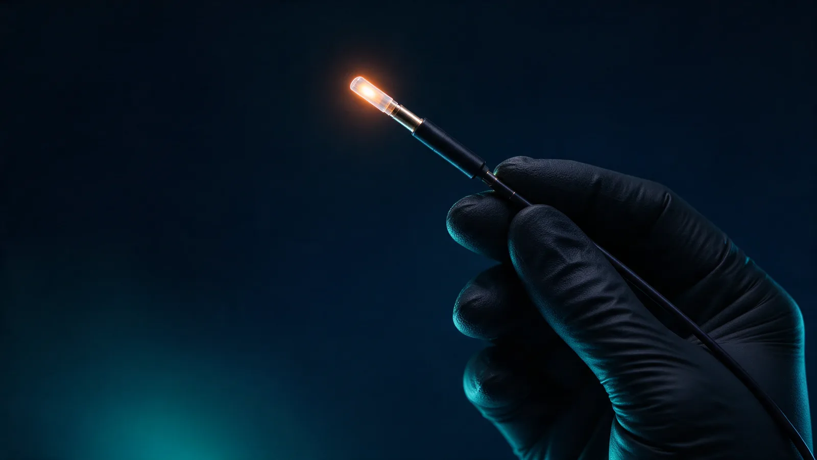
After allowing time for the drug to bind to cancer cells, a 690 nm red laser is applied 20–28 hours after the infusion ends. The light-activated dye damages the cancer-cell membrane, causing the cell to swell and burst. In the Japanese multicenter report, illumination was done under general anesthesia in the operating room, using one of two methods depending on the tumor shape.
- Surface illumination: light is applied from outside the tumor at 50 J per cm² for 5 minutes 33 seconds, with a 5–10 mm margin around the tumor edge.
- Interstitial illumination: thin needle catheters are placed into the tumor and cylindrical light fibers inserted through them deliver light from inside, at 100 J per cm of fiber for 4 minutes 10 seconds.
The technique keeps improving. In an analysis of a Japanese observational study of 77 patients at 24 centers, presented at the 2026 American Head & Neck Society (AHNS) meeting, covering the whole tumor with light at every session and using a narrower range per light source (5 mm depth for surface, 7 mm radius for interstitial) gave a volume-based complete response of 58.6%, compared with 42.9% for the wider setting. In the same study, the response rate per illuminated lesion was 60.8% and the disease control rate was 95.9%.
Step 4: Recovery and light care in hospital
After illumination, the treated area swells, and over several days the surface peels and tumor tissue sloughs away. Because the throat can swell, hospitals in the Japanese multicenter report secured the airway in advance: except for patients who already had a permanent tracheostoma or had nasopharyngeal cancer, they performed a preventive tracheotomy or intubation in the intensive care unit. The table shows the main side effects in this report (40 patients) and how they were managed.
| Side effect | All grades | Grade 3 or higher | Management |
|---|---|---|---|
| Pain at treated site | 92.5% | 12.5% | Controlled with pain medication |
| Mucositis | 80.0% | 7.5% | Mouth care and outpatient follow-up for 1–2 months |
| Bleeding | 77.5% | None | Wound observation and hemostasis |
| Laryngeal edema (throat swelling) | 42.5% | 10.0% (grade 4) | Airway secured by preventive tracheotomy or intubation |
Source: Okamoto I et al., Cancers 2025;17(16):2671. Grade 3 or higher side effects occurred in 17.5% overall, and 2 patients (5.0%) had sepsis, so infection control is also part of care.
Light care ends with a skin test. On day 6–7 after the infusion, a set amount of light is shone on the forearm to check for redness; if there is no reaction, the patient is discharged. In a Japanese study, none of 16 treatment sessions showed a photosensitivity reaction on this test, and no reaction appeared during one month after discharge. Patients are still advised to avoid direct sunlight for four weeks after the infusion. The average hospital stay was about two weeks, and outpatient follow-up continues for one to two months until mucositis settles.
Step 5: Checking the response and repeating if needed
After discharge, imaging every two to three months checks how the tumor has changed. If tumor remains, the same process can be repeated up to four times at intervals of at least four weeks (28 days). In the Japanese multicenter report, the 40 patients received 80 cycles in total, an average of two per patient.
In the Aichi Cancer Center study, most thin lesions reached complete response after a single cycle, while lesions of intermediate thickness reached complete response after two or more cycles. Complete response was more common when at least one interval between cycles was 50 days or less, so the researchers suggested scheduling the next session in advance according to tumor thickness.
| Study | Patients | Reported results |
|---|---|---|
| Phase 1/2a trial supporting approval (Study 101) | 30 patients with recurrent head and neck cancer | Response rate 43.3% (central review) |
| Japanese real-world care (Okamoto 2025) | 40 patients, 80 cycles | Response rate 75.0%, disease control 95.0%, median survival 26.9 months |
| Japanese observational study (AHNS 2026) | 77 patients at 24 centers | Per-lesion response rate 60.8%, disease control 95.9% |
| Aichi Cancer Center (Nishikawa 2025) | 19 patients, 30 cycles | Complete response in 7 of 14 local lesions |
Sources: Japan PMDA review report / Okamoto I et al., Cancers 2025 / Rakuten Medical AHNS 2026 presentation / Nishikawa D et al., Cancer Diagn Progn 2025. Patient groups and assessment methods differ, so the numbers are not directly compared.
What to prepare and questions for your doctor
- Medical records: details of previous surgery, radiotherapy and drug treatment, plus recent imaging
- Light protection items: long-sleeved clothes, gloves and a wide-brimmed hat (used until four weeks after the infusion)
- Schedule: about two weeks in hospital, outpatient visits for one to two months, and a stay plan that allows for repeat cycles
- Will my tumor be treated with surface illumination or interstitial illumination?
- Will I need a tracheotomy or intubation to protect my airway?
- How many cycles do you expect, and how will the interval be set?
- How long and how strictly should I avoid light after discharge?
Frequently asked questions
How long is the hospital stay for photoimmunotherapy?
In a Japanese multicenter report, the average hospital stay was about two weeks. Patients are discharged after passing the skin light test on day 6–7 and once wound recovery is confirmed; the length can vary with airway management and recovery speed.
How long should I avoid light after photoimmunotherapy?
In hospital, patients stay in a dimmed room of 80 lux or less until they pass the skin light test, and they avoid direct sunlight for four weeks after the infusion. Covering up with long sleeves, gloves and a hat is recommended over sunscreen.
How many times can photoimmunotherapy be given?
Up to four cycles can be given at intervals of at least four weeks. In a Japanese multicenter report patients received two cycles on average, and complete responses were reported after one cycle for thin tumors and after two or more for tumors of intermediate thickness.
Is photoimmunotherapy painful?
Laser illumination is done under general anesthesia, so no pain is felt during the procedure. Pain at the treated site afterwards was reported in 92.5% of patients and was mostly controlled with pain medication; grade 3 pain occurred in 12.5%.
Related: Head and Neck Cancer Photoimmunotherapy: Japan Approval and Results · Photoimmunotherapy Plus Pembrolizumab · Photodynamic Therapy vs Photoimmunotherapy · Photoimmunotherapy information
Sources
- PMDA (Pharmaceuticals and Medical Devices Agency, Japan) — Report on the Deliberation Results: Akalux (cetuximab sarotalocan sodium)
pmda.go.jp
Checked: 2026-10-30 - Okamoto I, Hasegawa O, Kushihashi Y et al. Real-World Effectiveness and Safety of Photoimmunotherapy for Head and Neck Cancer: A Multicenter Retrospective Study. Cancers 2025;17(16):2671
doi.org/10.3390/cancers17162671
Checked: 2026-10-30 - Nishikawa D, Shimabukuro T, Suzuki H et al. Predictive Factors for the Efficacy of Head and Neck Photoimmunotherapy and Optimization of Treatment Schedules. Cancer Diagn Progn 2025;5(2):179–188
cancerdiagnosisprognosis.org
Checked: 2026-10-30 - Yamakura T, Okamoto I, Okada T et al. Utility of a Simplified Irradiator for Photosensitivity Testing in Head and Neck Photoimmunotherapy. In Vivo 2026;40(4):2354
iv.iiarjournals.org
Checked: 2026-10-30 - Rakuten Medical — Investigation of Treatment Procedure to Optimize Photoimmunotherapy for Head and Neck Cancer (AHNS 2026 presentation, 2026-07-30)
rakuten-med.com
Checked: 2026-10-30
Written by the FTH editorial team. Sources checked 2026-10-30 · Medical review: not applicable
This article is general information based on public sources and does not replace diagnosis or treatment. Please discuss treatment decisions with your medical team.