Approved therapy — photoimmunotherapy laser illumination (approved in Japan in 2020, covered by national insurance since 2021): unresectable locally advanced or recurrent head and neck cancer · Results in this article: retrospective reports from Japanese university hospitals and the National Cancer Center (small, no control group)
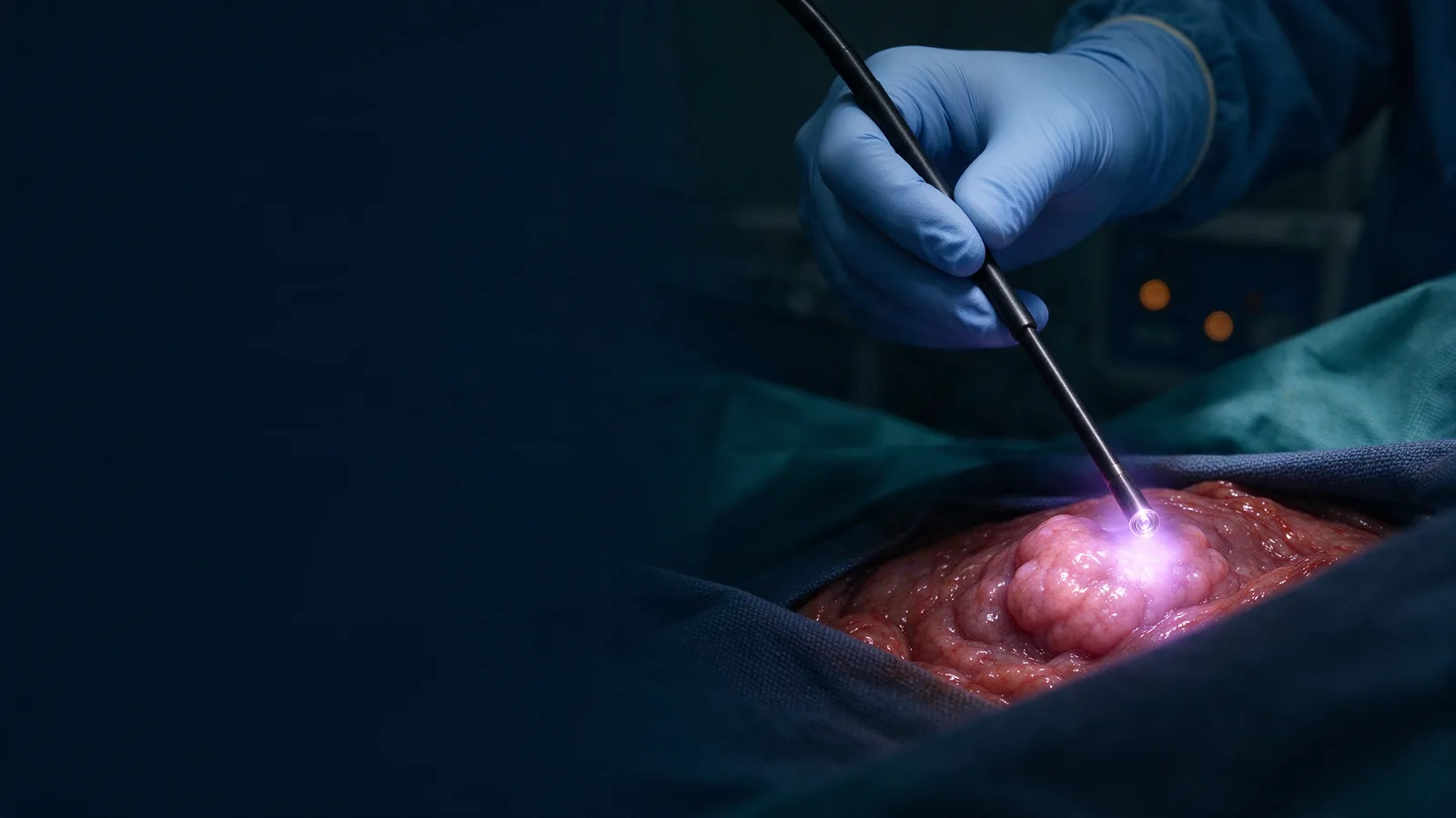
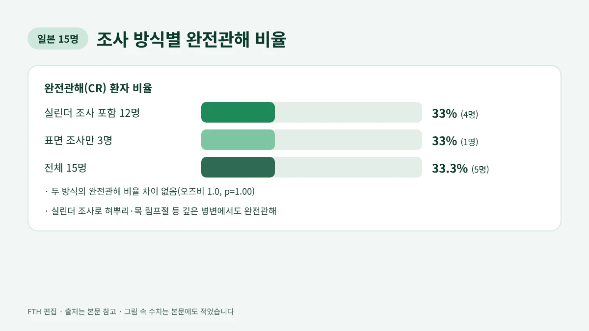
Photoimmunotherapy laser illumination is delivered either from the surface with a frontal diffuser or from inside the tumor with a cylindrical diffuser, depending on tumor thickness. In a 2026 report of 15 patients with recurrent head and neck cancer from the University of the Ryukyus in Japan, 4 of 12 patients treated with cylindrical illumination and 1 of 3 treated with surface illumination alone reached complete response, the same rate of 33% for both methods. Complete responses were reported even in deep-seated tumors treated with cylindrical illumination.
Why does photoimmunotherapy laser illumination use different methods?
Photoimmunotherapy uses a drug that binds to the EGFR protein on cancer cells (cetuximab sarotalocan, brand name Akalux), followed the next day by red laser light at 690 nm. The drug acts only where the light reaches, so delivering light to every part of the tumor strongly shapes the result.
The full sequence from infusion to discharge is covered in our photoimmunotherapy treatment process article, and overall outcomes in head and neck photoimmunotherapy results. This article collects only the results that Japanese university hospitals and the National Cancer Center have reported by illumination method.
Three photoimmunotherapy laser illumination devices and how they are chosen
The tip that releases light is called a diffuser, and Japanese hospitals choose it to fit the tumor. Based on reports from Hiroshima University (Tahara et al., Cancers 2026) and Kyoto Prefectural University of Medicine (Yoshizawa et al., Front Immunol 2026):
| Item | Surface | Side-fire | Cylindrical (interstitial) |
|---|---|---|---|
| Device | frontal diffuser | side-fire diffuser | cylindrical diffuser |
| Main use | Superficial tumors less than 1 cm thick | Superficial tumors hard to face directly | Tumors 1 cm thick or more |
| How light is emitted | Circular field from outside | Sideways | Radially from a fiber inside the tumor |
| Area per position | About 7–38 mm diameter | About 7–38 mm diameter | About 10 mm radius |
| Light dose | 50 J/cm² | 50 J/cm² | 100 J/cm of diffuser length |
Sources: Tahara et al., Cancers 2026;18(3):350; Yoshizawa et al., Front Immunol 2026;17:1814205. Hiroshima University illuminated 5–10 mm beyond the visible tumor edge.
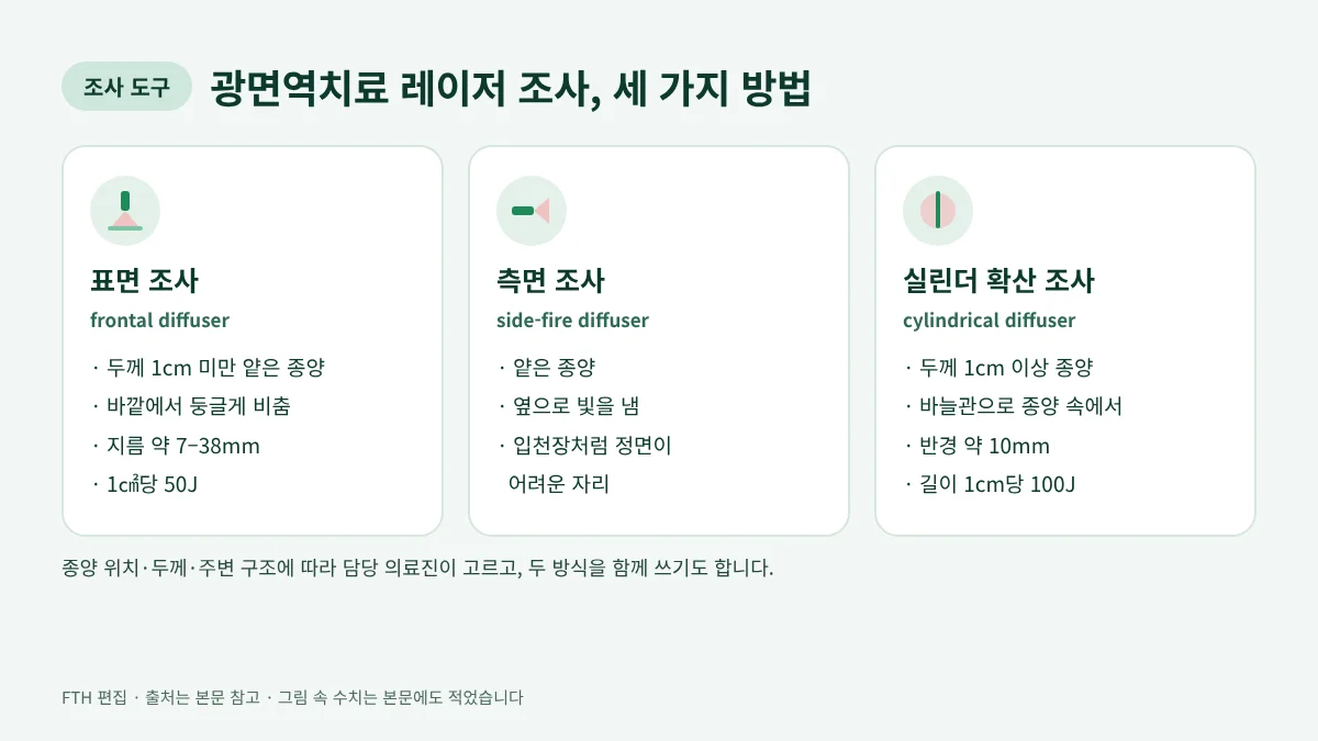
Hiroshima University showed the side-fire diffuser being used for a tumor of the soft palate. When one tumor has both shallow and deep parts, surface and cylindrical illumination can be combined in the same session.
Results by method: complete response with surface and cylindrical illumination
Researchers at the University of the Ryukyus treated 15 patients with recurrent head and neck squamous cell carcinoma who were not candidates for surgery, between January 2022 and December 2025. Using RECIST 1.1, 5 of 15 patients (33.3%) reached complete response, meaning no visible tumor on imaging (Hirakawa et al., Cancers 2026). The team published the device and outcome for each patient.
| Method | Patients | Complete response | Partial response |
|---|---|---|---|
| Cylindrical only | 9 | 4 | 3 |
| Cylindrical plus surface | 3 | 0 | 2 |
| Surface only | 3 | 1 | 0 |
| Total | 15 | 5 (33.3%) | 5 |
Source: Hirakawa et al., Cancers 2026;18(6):1022, Tables 2 and 3. Partial response means the tumor shrank by 30% or more.
Among the 12 patients who received cylindrical illumination (including 3 who also had surface illumination), 4 (33%) reached complete response, as did 1 of 3 (33%) treated with surface illumination alone. The team's statistical analysis found no difference in complete response by device (odds ratio 1.0, p=1.00). Complete responses occurred in the lateral oropharyngeal wall, base of tongue, floor of mouth and an anterior neck lymph node (all cylindrical) and in the nasopharynx (surface).
Deep tumors may seem hard for light to reach, but this report shows that placing fibers inside the tumor achieved the same complete response rate as in shallow tumors. Because this is a 15-patient study without a control group, the rate is a reference value to be confirmed in larger studies. In the same study, patients with a lower pretreatment inflammation index (SIRI) more often reached complete response (median 70.7 vs 120.2, p=0.03).
Catheter spacing matters for cylindrical illumination
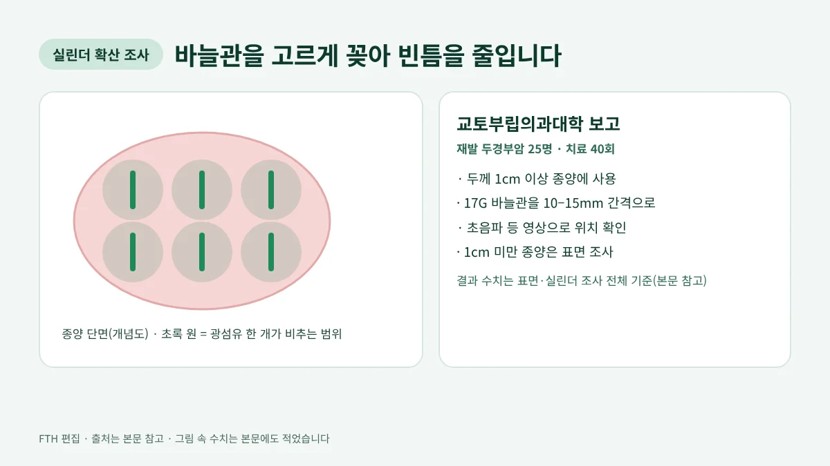
For cylindrical illumination, thin needle catheters are inserted into the tumor and a light fiber is placed inside each one. Each fiber covers a radius of about 10 mm, so several must be spread evenly to cover the whole tumor.
Kyoto Prefectural University of Medicine inserted 17-gauge catheters at 10–15 mm intervals into tumors 1 cm thick or more, checking their position with imaging such as ultrasound, and used surface illumination for tumors under 1 cm. Across all 25 patients with recurrent head and neck squamous cell carcinoma treated this way over 40 cycles, with both methods matched to the tumor, the response rate was 77.5% (7 complete and 24 partial responses) and median overall survival was 401 days. The main adverse events were local reactions such as swelling and pain (grade 3 edema in 6 instances), and 11 patients (44%) had a planned tracheostomy beforehand to protect the airway, as done for oropharyngeal and hypopharyngeal lesions (Yoshizawa et al., Front Immunol 2026).
An analysis of 77 patients at 24 Japanese centers, presented at the American Head and Neck Society meeting in 2026, also found more complete responses when each light field was set smaller and the tumor was covered without gaps (Rakuten Medical, 2026-07-30). Those figures are in step 3 of our treatment process article.
How do pain and recovery differ by illumination method?
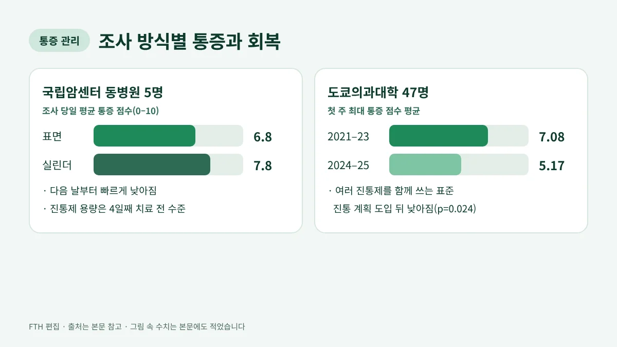
Cylindrical illumination, which requires needle catheters, is somewhat more invasive than surface illumination. A case series of 5 patients treated at National Cancer Center Hospital East from January 2021 to June 2022 (Shibutani et al., Healthcare 2023) found mean pain scores (0–10) on the day of illumination of 6.8 with surface and 7.8 with cylindrical illumination.
Pain peaked right after or one hour after illumination and eased quickly the next day. All patients treated with cylindrical illumination had their pain controlled with opioids, and the opioid dose returned to the pretreatment level by day 4. One of the two patients treated with surface illumination did not need opioids.
Pain management is improving. Tokyo Medical University reported that after a standardized multimodal analgesic plan was applied, mean maximum pain in the first week fell from 7.08 in 24 patients treated in 2021–2023 to 5.17 in 23 patients treated in 2024–2025 (p=0.024, Yoshida et al., In Vivo 2026). Tumor size was not clearly related to pain intensity.
Techniques for reaching deep, narrow sites
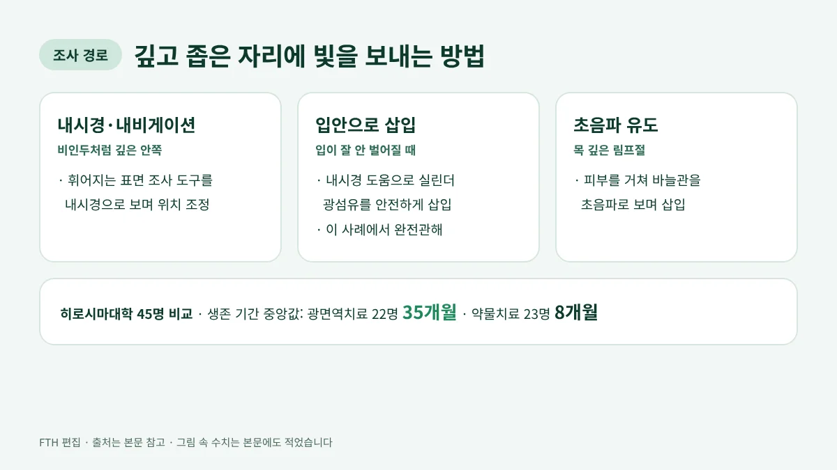
The head and neck has tight spaces and large blood vessels nearby, so techniques for guiding light are advancing too. The University of the Ryukyus report described these examples:
- Nasopharyngeal cancer: a flexible frontal diffuser was guided to the nasopharynx under nasal endoscopy and a navigation system.
- Oral tumor in a patient who could barely open the mouth: a cylindrical fiber was inserted through the mouth with endoscopic help, and this patient reached complete response.
- Deep neck lymph node: catheters were inserted through the skin under ultrasound guidance.
Hiroshima University compared 45 patients with unresectable head and neck cancer without distant metastasis treated between January 2021 and April 2025. Median overall survival was 35 months in the 22 patients who received photoimmunotherapy versus 8 months in the 23 who received drug therapy. In a multivariable analysis limited to patients eligible for photoimmunotherapy, it remained associated with longer survival (hazard ratio 0.24, Tahara et al., Cancers 2026). As a single-center retrospective study, the authors called for multicenter prospective studies to build on it.
Japanese multicenter results for the nasopharynx are in nasopharyngeal cancer photoimmunotherapy, and approval scope and basics in photoimmunotherapy information.
Questions for your doctor
- How thick is my tumor in mm, and is surface or cylindrical illumination suitable?
- If cylindrical, how many catheters will be used and what imaging guides them?
- How wide a margin beyond the tumor edge will be illuminated?
- What pain plan is used on the day of illumination?
- If tumor remains after one cycle, when would the next illumination be?
Frequently asked questions
Which works better, surface or cylindrical illumination?
In the 15-patient comparison from the University of the Ryukyus, the complete response rate was the same, 33%, for both. The method is chosen to fit tumor thickness and location rather than by effectiveness, and the care team makes that decision.
How long does photoimmunotherapy laser illumination take?
It varies by patient, because larger tumors are illuminated in several positions. Time per position and the full schedule are in our treatment process article.
Is cylindrical illumination more painful?
In the 5-patient series at National Cancer Center Hospital East, mean pain on the day of illumination was 7.8 with cylindrical and 6.8 with surface illumination. Pain was controlled with medication, and the opioid dose returned to the pretreatment level by day 4.
How many times can laser illumination be repeated?
Of the 15 patients at the University of the Ryukyus, 7 received a single cycle, and 3 of them reached complete response. Repeat intervals and the maximum number of cycles are in our treatment process article.
Sources
- Hirakawa H, Ikegami T, Kinjyo H, et al. Nutritional and inflammatory markers associated with complete response to near-infrared photoimmunotherapy in recurrent head and neck squamous cell carcinoma. Cancers 2026;18(6):1022
doi.org/10.3390/cancers18061022
Checked: 2026-11-19 - Tahara H, Ueda T, Hattori T, et al. Outcomes of near-infrared photoimmunotherapy for head and neck cancer: a single-center retrospective study. Cancers 2026;18(3):350
doi.org/10.3390/cancers18030350
Checked: 2026-11-19 - Yoshizawa K, Tsujikawa T, et al. EGFR-targeted photoimmunotherapy and its association with the immune microenvironment in locoregional recurrent head and neck squamous cell carcinoma. Front Immunol 2026;17:1814205
doi.org/10.3389/fimmu.2026.1814205
Checked: 2026-11-19 - Shibutani Y, Sato H, Suzuki S, et al. A case series on pain accompanying photoimmunotherapy for head and neck cancer. Healthcare 2023;11(6):924
doi.org/10.3390/healthcare11060924
Checked: 2026-11-19 - Yoshida S, Okamoto I, Tokashiki K, Tsukahara K. Pain associated with head and neck cancer photoimmunotherapy. In Vivo 2026;40(5):3014–3022
doi.org/10.21873/invivo.14450
Checked: 2026-11-19 - Rakuten Medical — Investigation of treatment procedure to optimize photoimmunotherapy for head and neck cancer (AHNS 2026, 2026-07-30)
rakuten-med.com
Checked: 2026-11-19
Written by the FTH editorial team, sources checked 2026-11-19 · Medical review: none
This article is general information based on public sources and does not replace diagnosis or treatment. Please discuss treatment decisions with your care team.